Floor Of Sella Turcica Bone
Floor Of Sella Turica Definition Of Floor Of Sella Turica By Medical Dictionary

Sellar Parasellar And Clival Region Radiology Key

Specialised Projections Of The Skull Radiology Key
Middle Cranial Fossa Boundaries Contents Teachmeanatomy
Sella Turcica And Pituitary Gland Radiology Key
Solved 14 15 17 18 15 20 21 22 23 24 10 25 11 26 12 A S Chegg Com

Miller keane encyclopedia and dictionary of medicine nursing and.
Floor of sella turcica bone.

Anatomy Ch 07 Cranial Floor Images Flashcards Quizlet

Sphenoid Bone Chromoscience

Keptalalat A Kovetkezore Sella Turcica Anatomy Bones Axial Skeleton Medical Anatomy
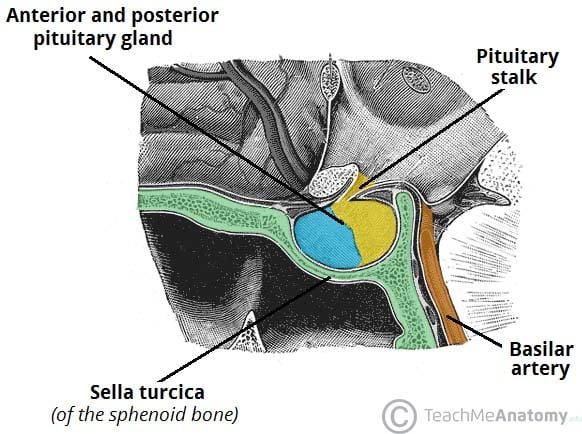
The Pituitary Gland Structure Vasculature Teachmeanatomy
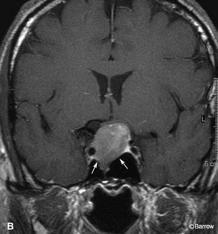
Double Sellar Floor Radiographic Sign For A Pituitary Adenoma Barrow

Sella Turcica Image Yousun Koh Sphenoid Bone Bones Sinusitis

The Sensory Nerves Chromoscience

The Skull Bone And Features Flashcards Quizlet

Enlarged Sella Turcica Differential Radiology Reference Article Radiopaedia Org

Sella Turcica An Overview Sciencedirect Topics
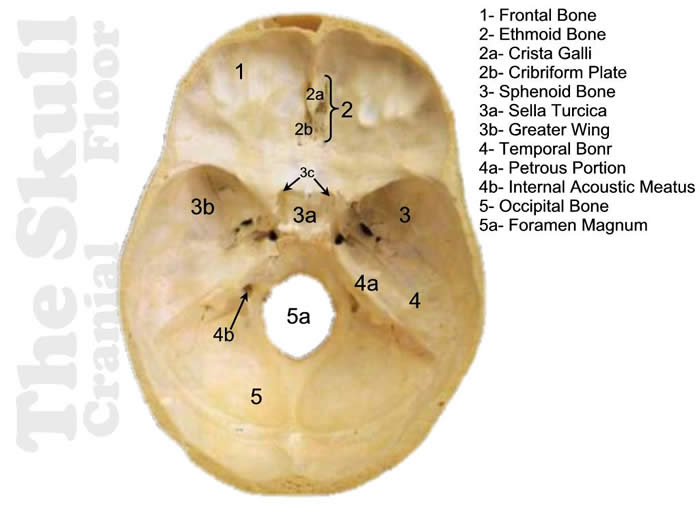
Dem Bones Skull Cranial Floor Answers
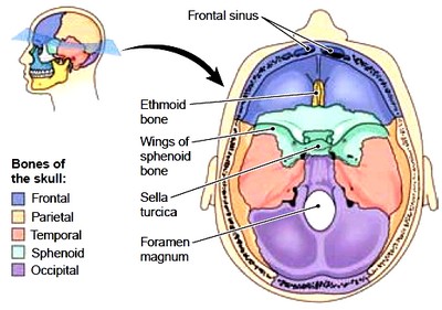
Axial Skeleton Cranium Facial Bones Vertebral Column Thorax

Skeletal Sysyem Module 8 The Skull Skeletal System Openstax Cnx

Sella Turcica Image Yousun Koh Sphenoid Bone Anatomy Bones Skull Anatomy

Ch 9 Spenoid Bone Flashcards Quizlet
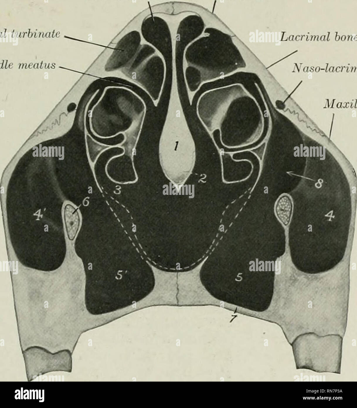
The Anatomy Of The Domestic Animals Veterinary Anatomy Skull Of The Ox As A Whole 143 Than The Rest Of The Floor The Ethmoidal Fossffi Are Smaller And The Hypophyseal Fossa

A P 106 Chapter 7 Axial Skeleton Flashcards Quizlet
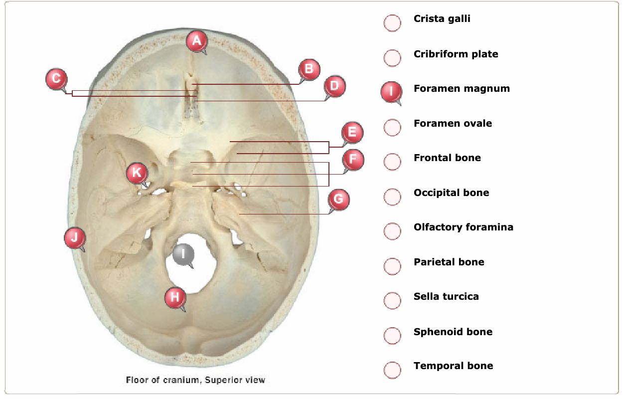
Solved Crista Galli Cribriform Plate S Foramen Magnum For Chegg Com
Https Encrypted Tbn0 Gstatic Com Images Q Tbn 3aand9gcqnfu7kj9hqxes7vatdmueowu0z9osltbm2nmjyzewytwmfthet Usqp Cau

Slides Show
The Base Of The Skull Superior View Of Cranial Floor Anatomy Images Illustrations Anatomy Images Charac Craniosacral Therapy Anatomy Anatomy Coloring Book
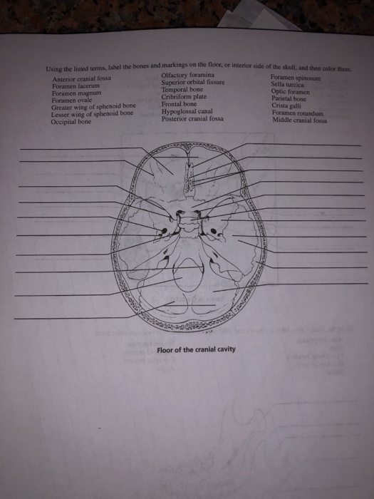
Solved On The Floor Or Interior Side Of The Skull And T Chegg Com
Https Link Springer Com Content Pdf 10 1007 2f978 3 642 67786 1 13 Pdf

Full Text Association Of Sella Turcica Bridging With Palatal Canine Impaction In Ccide
Source : pinterest.com
