Floor Of Posterior Triangle Of Neck Is Formed By
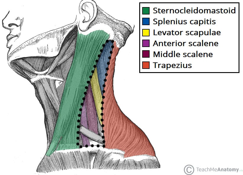
Posterior Triangle Of The Neck Subdivisions Teachmeanatomy
Easy Notes On Posterior Triangle Of The Neck Learn In Just 3 Mins Earth S Lab

Posterior Triangle Of Neck Boundaries Contents Sternocleidomastoid Muscle Anatomyqa
Posterior Triangle Of The Neck

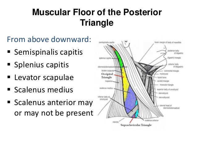
Occipital Triangle Wikipedia

Zoology Human Neck
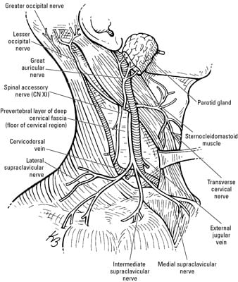
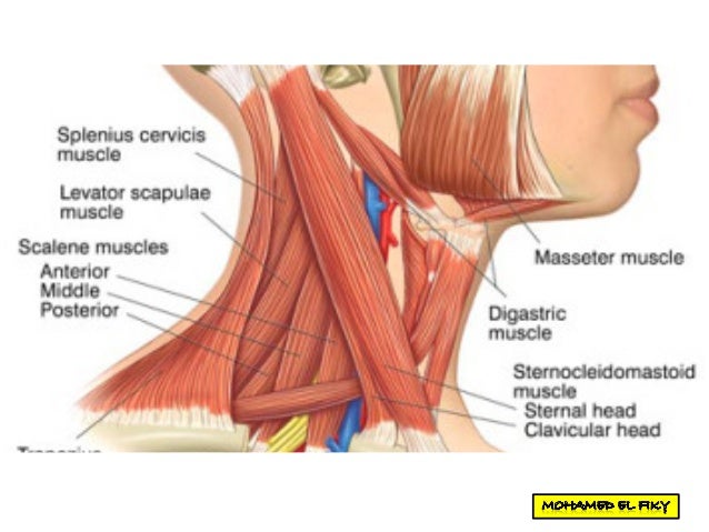
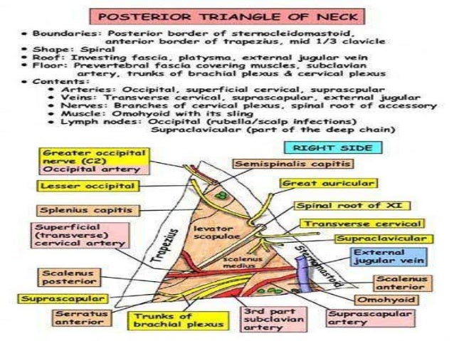
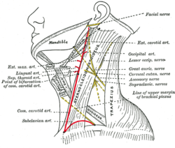
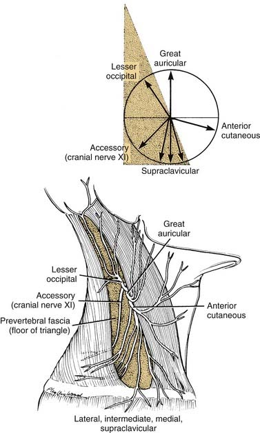
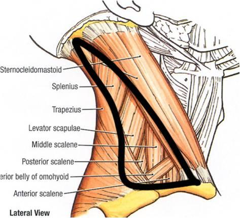
It s about the posterior triangle of the neck today.
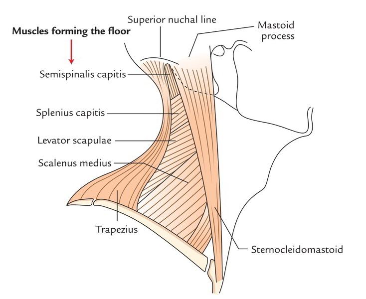
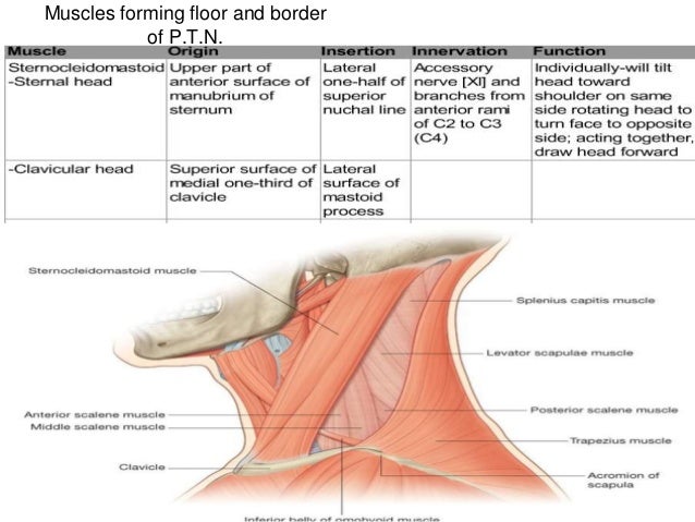
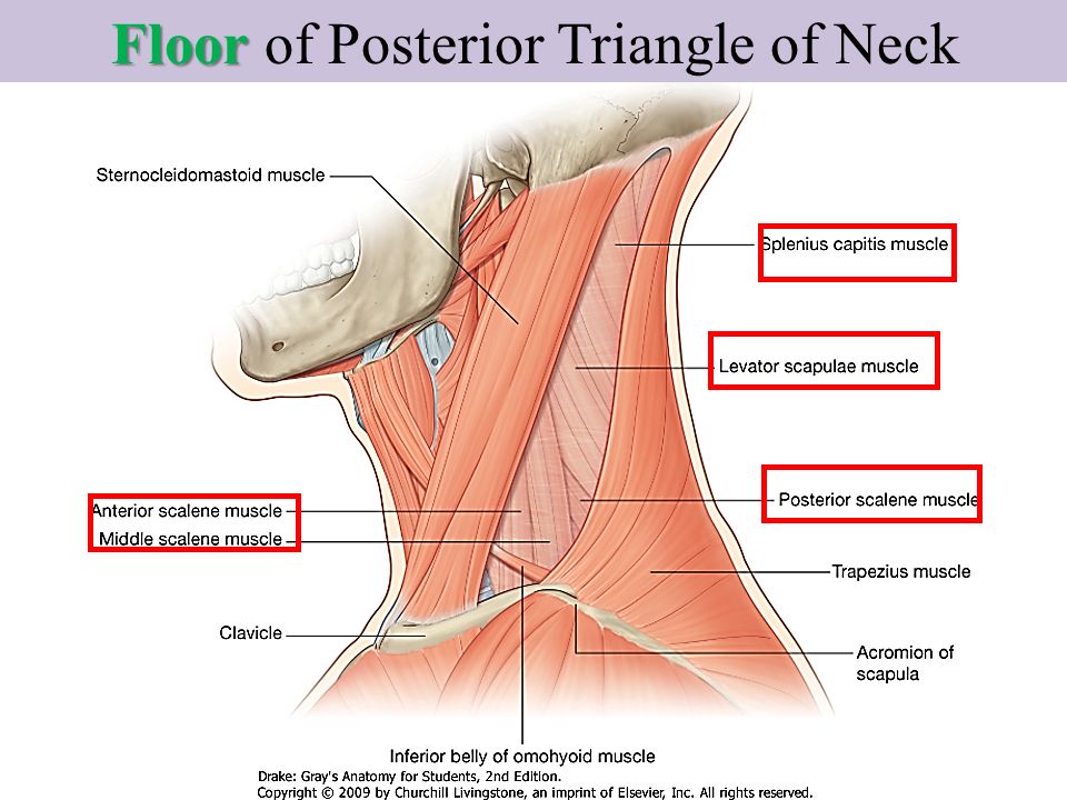
Floor of posterior triangle of neck is formed by.
Posterior Triangle Of The Neck

Neck Fascial Layers Posterior Triangle Anterior Triangle Trachea Thyroid Flashcards Quizlet
Neck Triangles Lecture 3 Anatomy Block 2 Flashcards Memorang
Posterior Triangle Of Neck
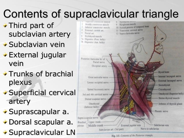
Subclavian Triangle Wikipedia
Posterior Triangle Of Neck

Posterior Triangle Of The Neck Everything You Need To Know Dr Nabil Ebraheim Youtube
Scalp Muscles Of Face D Rania Gabr D Sama D Elsherbiny Ppt Download

Posterior Triangle Of Neck Www Medicoapps Org

Triangles Of The Neck Anatomy Borders And Contents Kenhub

Image Result For Posterior Triangle Of Neck Medical Anatomy Brachial Subclavian Artery

Floor Of The Posterior Triangle Anatomy Mitch Medical Healthcare

Image Result For Posterior Triangle Of Neck Borders Triangle Borders Neck

Image Result For Posterior Triangle Contents Subclavian Artery Skeleton Anatomy Triangle

Triangles Of The Neck A Review With Clinical Surgical Applications Abstract Europe Pmc
Anatomy And Developmental Embryology Of The Neck Ento Key
The Posterior Triangle Of The Neck Dummies

Head Neck Unit Lecture 11 د حيدر جليل الأعسم Ppt Video Online Download
1
Triangles Of The Neck Part 1 The Anterior Triangle Medical Exam Prep
001b Superficial And Deep Structures Of The Neck Anatomy Ii Flashcards Memorang
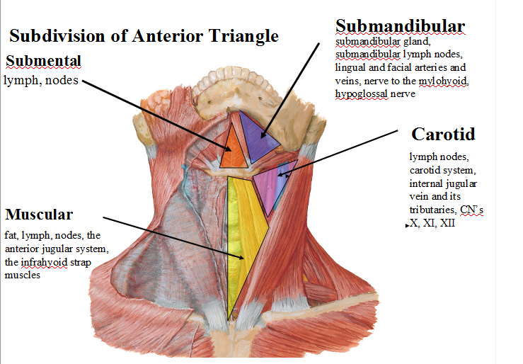
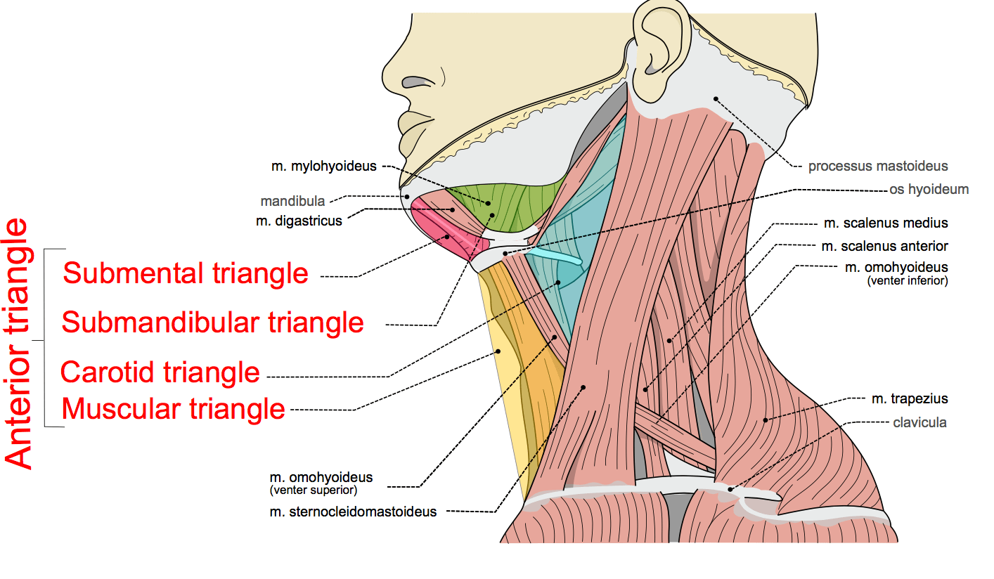
Anterior Triangle Of The Neck Subdivisions Teachmeanatomy

Cb 724 Triangles Of The Head And Neck Flashcards Quizlet
Posterior Triangle Of Neck
Source : pinterest.com
